
My Dutch Results
There is a saying in finance that “what you don’t measure you cannot improve” and I think that this holds true for our health as
There is a saying in finance that “what you don’t measure you cannot improve” and I think that this holds true for our health as

back to all Since vitiligo is an autoimmune disease, certain dietary patterns and supplements may be used to support the immune system in individuals with

In most people’s minds breath holding is synonymous with dysfunctional breathing. Despite its reputation the benefits of voluntary breath holds are far reaching and can

Breathwork, by changing our blood’s biochemistry, can influence the function of all body’s organs and systems. As oxygen is essential for aerobic respiration, and all

It is not uncommon for runners to gas out when running. Their lungs will fail them before their legs. While this is more common among
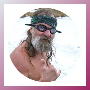
Those practicing the Wim Hof Method will benefit primarily from the progressive exposure to cold. The regular cold showers the method advocates will: • up

Breathwork is one of the most powerful tools in dealing with stress and anxiety. Scientific research and clinical evidence have proven that 4-6 weeks of

Developed by Russian doctor Konstantin Buteyko in the 1950s the Buteyko Method is a system aiming to restore the levels of healthy CO2 in the body

Leaky gut is the result of structural damage in the intestines. With the gut permeability been compromised, most individuals experience a series of symptoms, not