Leaky gut is the result of structural damage in the intestines. With the gut permeability been compromised, most individuals experience a series of symptoms, not solely digestive. To heal leaky gut permanently a structured multidisciplinary approach is needed.
Leaky gut: is a digestive track with a compromised permeability (like a hose with holes).
.
The Intestinal Immune System
The reason why leaky gut symptoms are not exclusively digestive are due to gut’s double role:
• digest and absorb nutrients
• host part of the immune system
The immune system in the gut has the delicate role of balancing between: Tolerating or Reacting to the foods it comes in contact with. The evolutionary benefit of this role is the following:
Foods we consume may be degraded or containing toxins and thus be poisonous to the body. In these cases the activation of the immune system can kill the pathogenic substances and protect us. This process is mediated through a series of steps leading to the increase of intestinal permeability.
Unfortunately in certain people the same reaction is triggered not only by toxins but also by regular foods. In these cases after the consumption of the “trigger food” the individual experiences a reaction such as: foggy brain, bloatedness, diarrhea, stomach cramps, increased heart rate, running nose, anxiety, irritability. Reducing gut permeability (i.e. healing leaky gut) can make previous “trigger foods” tolerable again.
How do you test for leaky gut?
Our gut wall consists of just one cell thick epithelial tissue (Sturgeon, C. and Fasano, A., 2016). The space between each epithelial cell is called tight junction. The following tests can be used to identify leaky gut.
.
Lactulose/Mannitol test
The test that has been used the longest for detecting leaky gut is the lactulose/mannitol urine test. The test is simple: after an overnight (12 hour) fast you collect the urine then have a solution of lactulose & mannitol and 6 hours later you collect the urine again.
Mannitol enters the body through the epithelial cell membrane, while lactulose goes through the tight junctions (FlemIng, S.C. et al., 1990)
The loss of absorptive areas ➛ ↓ the absorption of mannitol.
The loss of mucosal integrity ➛ ↑ lactulose absorption.
.
An elevated lactulose: mannitol ratio indicates the presence of leaky gut. The test is available from many labs including Genova Diagnostics. The results can be affected by the use of NSAIDS, alcohol and according to Dr. Alesio Fasano the results are very sensitive to the collection process and thus may not be reliable when done outside a lab.
.
3 stool markers of leaky gut (α1-Antitrypsin – sIgA – calprotectin)
α1-Antitrypsin
is a protein of the liver. When detected in stool (sourced from the intestines) it indicates a severe case of intestinal permeability and thus is not a sensitive enough marker of leaky gut (Biancone, L. et al., 2003)
sIgA
is part of the immune system and functions as a tag for substances that need to be excreted.
Calprotectin
is a protein linked with intestinal inflammation. It is used to distinguish between IBD & IBS (Leblhuber, F., et al., 2015).
.
3 blood markers of leaky gut (Zonulin – LPS – DAO)
Zonulin
Zolulin is a protein responsible for the modulation of tight junctions (Sturgeon, C. and Fasano, A., 2016).
↑ levels of Zonulin ➛ Opening of tight junctions ➛ influx of dietary & microbial antigens in the blood
The 2 main triggers of Zonulin release have been found to be:
- Bacteria: including Eschericha coli, lab E. coli, virulent E. coli, and Salmonella typhi (El Asmar et al. 2002)
- Gliadin: a protein found in gluten (Clemente, M et al., 2003)
Elevated levels of Zonulin have been linked in the literature (Sturgeon, C. and Fasano, A., 2016) to:
- autoimmune conditions such as Type 1 Diabetes, Celiac Disease, Multiple Sclerosis, Intestinal Bowel Diseases
- metabolic disorders such as: Obesity & PCOS
- Asthma
- Coronary Heart Disease
- Systemic infections
- Gluten Sensitivity
- Necrotizing Enterocolitis
- Brain cancer (Skardelly, M et al., 2009) by altering the integrity of the Blood Brain Barrier.
.
Lipopolysaccharide Bacterial Endotoxin
Lipopolysaccharide (LPS) is a component of the wall of gram-negative bacteria (Trent, M.S et al., 2006) responsible for the activation of the innate immune system. LPS has 3 regions. Lab tests measure the lipid A region which is also known as endotoxin. Germ-negative bacteria live in the lumen of the gut but should not be found in the blood. Detection of LPS endotoxins in the blood is a sign of leaky gut.
.
DAO
Dunwoody Labs measures the levels of DAO enzyme in their intestinal permeability test. DAO is responsible for the break down of histamine. Histamine while necessary for good gut health when elevated can cause problems. Low levels of DAO thus is also a sign of leaky gut. Genetic polymorphisms in the AOC1 gene (which encodes the DAO enzyme) can impair the body’s ability to produce the DAO. Those with low levels can check their genetic burden using the table below.
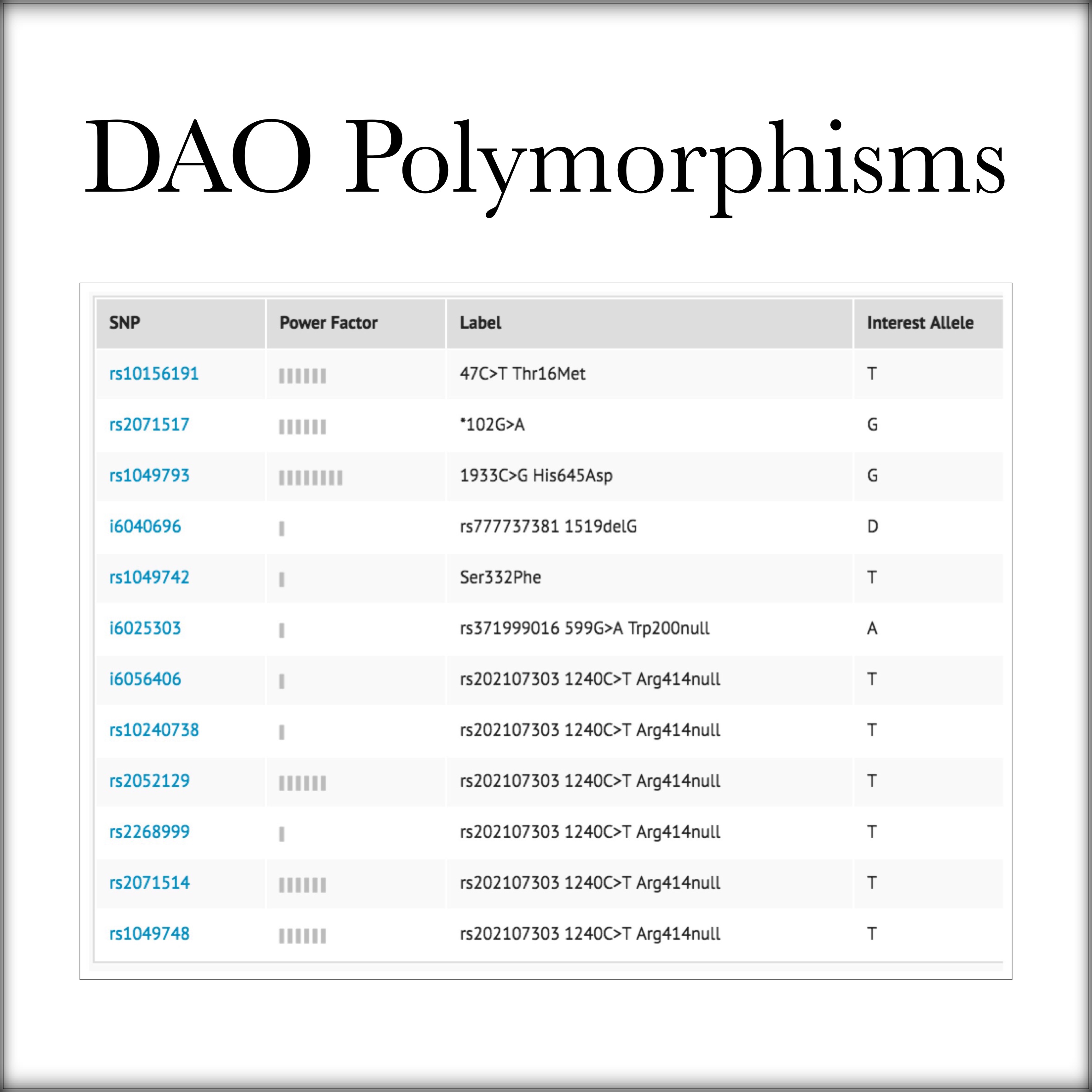
source: Opus23
.
The future of leaky gut testing
While not currently available for the general public I-FABP is a marker of gut permeability used in laboratories. Intestinal fatty acid binding protein (I-FABP), is a marker of early enterocyte cell death (Derikx, J.P. et al., 2010)
.
How to support a leaky gut?
When it comes to supporting leaky gut I like to split the nutrients in 2 categories:
- the ones affecting the mechanisms that cause the problem (these are the ones that ultimately will heal the intestines)
- the ones that suppress the symptoms – commonly referred to as anti-inflammatory (these are the ones that should help ameliorate the symptoms)
.
Avoid trigger foods
While I consider elimination diets not a good idea long-term in the short run it is important to remove any trigger foods to control inflammation. IgG food intolerance tests can be very useful for that matter.
.
Probiotics
I consider the use of probiotics the most potent yet the most tricky in implementation among all interventions. Certain probiotic strains have been found to induce cell proliferation in gut cells:
Bifidobacterium breve R0070 & Lactococcus lactis R1058, when taken together, seem to have synergistic effects and both can be found in the Jarrow-Dophilus EPS. (Grimoud, J. et al., 2010 *)
Some others that were shown to suppress inflammation induced by LPS levels are:
- Bifidobacterium longum subsp. Infantis, Bifidobacterium longum, Bifidobacterium bifidum, Lactobacillus rhamnosus – in the order mentioned (Laetitia, R. et al., 2013)
- Lactobacillus reuteri strain, ATCC PTA 6475 – available from Biogaia. (Thomas, C.M. and Versalovic, J., 2010)
- Bifidobacterium infantis 35624 (Groeger, D. et al., 2013)
.
* The French study by Julien Grimoud is a goldmine of information.
.
Mushrooms
Berberine
Berberine is an alkaloid found in some plants shown to inhibit the inflammatory effects of LPS (Mo, C. et al., 2014, Wu, Y.H., et al., 2012). Berberine has been shown to interact with 57 genes, so cross-checking polymorphisms related to other symptoms is worth doing.
.
Other agents
Quercetin & CoQ10 were also shown to have anti-inflammatory effects in LPS toxicity (Abd el-gawad, H.M. and Khalifa, A.E., 2001). Fish Oils were shown to restore intestinal integrity by increasing DAO enzyme concentration in the gut (Liu, Y. et al., 2012).
.
L-glutamine
L-glutamine acts as fuel for intestinal cells (Larson, S.D, et al., 2007), and to that extent supplementation can benefit a leaky gut. Gradually building the dosage from as little as 2.5 gr per day to 20 gr should be a safe way to avoid adverse reactions. I have not seen any studies demonstrating the benefits of L-glutamine supplementation for leaky gut however it does support overall intestinal health.
.
Larazotide acetate
Larazotide acetate is a protein shown to inhibit Zonulin production without any adverse effects (Paterson, B.M et al., 2007). Alba Therapeutics an Indian pharmaceutical company is in the process of developing a drug with this protein.
Frequently Asked Questions
Is Fodmap diet good for leaky gut?
Many individuals suffering from leaky gut, experience food sensitivities. The easiest way to deal with food sensitivities is to adopt an elimination diet. While this initially is a good idea as inflammation goes down, long term they cause problems.
.
That’s why in my opinion:
- While elimination diets (i.e. FODMAP, low oxalate, low histamine diets) remove foods that cause reactions, the reason why the reaction was there are 1st place stays. The same foods that
- Most chronically ill patients have restricted diets: the body is not able to renew the epithelial tissue in the gut leading to poor gut integrity ➛ increased gut permeability ➛ food sensitivities.
- Diversity in gut flora is positively associated with health: A diverse gut flora supports gut integrity.
.
Can Candida cause Leaky Gut?
Candida can cause intestinal imbalances and sequentially leaky gut. As candida symptoms overlap with other GI tract issues it is worth testing for candida prior to following a protocol.
.


.
References
20 responses